דף הבית » Computerized Total-Body Photography – Mole Mapping
Background
Digital mole mapping, also referred to as Total-body photography, complements the total-skin examinations.
The test is based on a simple principle: benign skin lesions generally remain stable over time, while malignant tumors grow and change.
Whenever you visit your dermatologist, even if it is a follow-up appointment, the examination is conducted as if it were your first encounter, because even the most skilled dermatologists cannot remember the appearance of each and every lesion from six months or a year ago. Therefore, a mole examination focuses on the appearance of lesions at a particular moment and does not consider their behavior over time. This is where mole mapping comes into play, addressing this limitation.
How is the test performed?
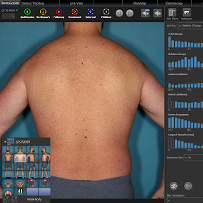
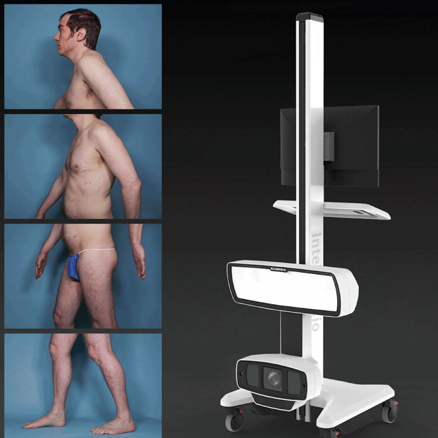
Digital mole mapping entails capturing photographs of the patient’s entire skin surface area. A technician or nurse operates a semi-automatic device, and the patient assumes different positions in front of a camera mounted on a rail. The camera moves up and down, capturing images of the entire skin surface. These images are stitched together to create a comprehensive “map” documenting the patient’s entire skin area. A year later, the imaging process is repeated to generate an updated “map” for comparison with the previous one. By comparing the two maps, even subtle changes in the patient’s lesions can be detected, allowing for the early identification of suspicious lesions that might not be apparent during a regular mole examination. Some advanced systems even incorporate artificial intelligence technology to assist doctors in comparing the maps and identifying changing and suspicious lesions.
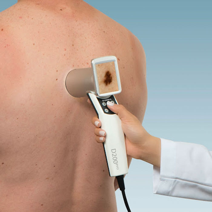
Digital dermoscopy is another component of mole mapping. This technique involves taking close-up images of suspicious skin lesions using a dermoscopic camera. These close-up images are revisited three to four months later and compared to the initial images. If microscopic changes are observed in the lesion, it becomes a suspicious lesion requiring a biopsy. Conversely, if the lesion remains stable, follow-up can be conducted. Digital dermoscopy helps avoid unnecessary biopsies, as lesions that appear highly suspicious during initial examination but remain stable over time are determined to be benign and can be safely monitored.
Additional relevant information
Digital mole mapping is a non-invasive test that does not involve radiation exposure or any discomfort. It is free of side effects.
This test is frequently recommended for individuals at an elevated risk of skin cancer, including those with a personal or family history of the disease, multiple moles, sun-damaged skin, excessive sun exposure history, and more.
Currently, the test is not covered by the public health insurance in Israel and is offered as a fee-based service. Depending on the patient’s insurance policy, reimbursement for the test may be possible through private medical insurance.
In Conclusion
If you have located a suspicious lesion on the skin, especially if it is unusual, new or recently changed, contact a dermatologist as soon as possible in order to diagnose the lesion and decide on further treatment.